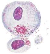
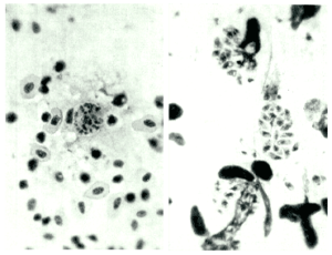
Cervical smear infected with RickettsiaA few decades ago, rickettsia were consecutively considered as being viruses or bacteria. In fact they are neither.
They belong to a group of intracellular germs with a poor enzymatic component. Therefore, they have to use for survival, the ensymatic system of the cell they
invade. This is why we call them obligated intracellular organisms.
A disease called Fatigue – p 153
This infection can present as an acquired or congenital form, acute or chronic, generalised or localised. In the brain, heart, skin, eyes, glands. Anywhere its
evolution can be slow, in the shelter of a dark cyst. It can cause abortion or severe visual impairment in the newborn.
Today this serious disease is widespread and, despite intensive researches following its fortuitous discovery, the treatment is difficult, relapses are
frequent and very often the diagnosis is missed.
A disease called Fatigue – p 31
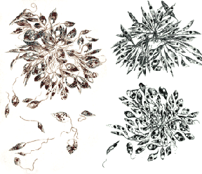
Another species of the Trypanosomatida family. Also adapted to the intracellular life in human kind. It has been identified in Europe, Asia, Africa, and from Mexico to Argentina and Chile. Parasiting the liver, the spleen, the bone marrow and the lymph nodes of the endothelial tissue in case of visceral leishmaniosis. In this cases, the IgE will drastically increase. Anaemia will be detected as well as splenomegaly and repeated bouts of fever. Another important form of Leishmaniosis is the so called “Bouton d’Orient” and other ulcerations where the germ can stay alive for more than twenty years in the subcutaneous tissue.
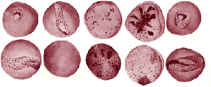
Malaria remains a fierce killer of humankind. It kills between one and three million people every year – half a holocaust per annum.
A disease called Fatigue – p 38
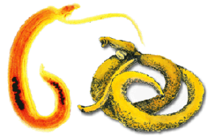
Even though Bilharzia is one of the prominent diseases in South Africa, it often remains undiagnosed, as the expected blood in the urines is a rather rare
symptom testifying the presence of the worm.
This parasitic disease is caused by a Trematode (blood flukes)of the genus Schistosoma.
Bilharzia infects about 200 million people worldwide, put 600 millions at risk and kills 250 thousand annually.
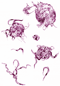
One important species of the Trypanosomatida family. Trypanosoma brucei gambiense is transmitted by hanging onto the proboscis of the tsetse fly. This fly moves very little, lives in some areas of Africa and is a glutton, feeding every day. Trypanosoma brucei rhodesiense is an acute form of the sleeping sickness. This disease expresses with a sudden laziness, the face of a greedy guinea pig and the neck of an ostrich, which seems to have swallowed a few jewels to testify to the passage of tourists. This is caused by cervical gland hypertrophy. This protozoa takes up residence preferably in humans or in dogs, but also in camels, pigs, cows, goats and sheep. It is principally an African disease, found in endemic and epidemic forms. Its victims are thus far a medical novelty. It is found from Central Africa to the Zimbabwean borders, and in Latin America. My father managed to isolate it from the blood of bats that were haunting our attic in Antwerp. How often has this condition been misdiagnosed as the popular Glandular Fever – Ebstein Barr virus?
A disease called Fatigue – p 35-36
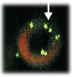
Section of human red blood cell infected with Bartonella quintana. Bartonelle quintana is transmitted by body lice. Infection with this organism leads to chronic bacteraemia with few symptoms.
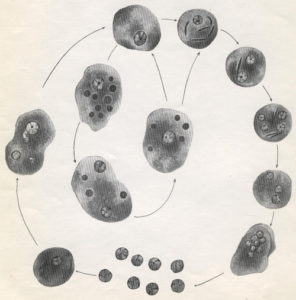
Found in contaminated water. It can cause digestive problems, livers’ abscess, lungs’ abscess and meningitis or encephalitis. The search of cysts with four
nucleus in the stool of patients shows that the parasite is cosmopolitan.
Due to its size, it could be compared to a large cruiser where smaller germs take place and even survive there after the elimination of the amoeba from the
human body.
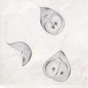
1. Intestinalis: Parasites the intestine where it feeds by osmosis- real leaches on intestinal mucosa. It has a round structure that enables it to do so.
As a consequence, the carrier will experience diarrhoea with flatulence followed by constipation. This will also produce ulcerations and malabsorption.
They are know to invade the gallbladder or the mouth and the stomach.
2. Vaginalis: To be found in the vagina, the urethras and the prostates. It cannot be cultivated in alkaline solution but only in acid environment. It will
do better where endocrines abnormalities are present.
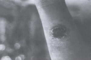
Leishmanian ulcer with elevated edges where parasites will be found. The middle becomes progressively scar tissue and contains no parasites.
The causative agent is Francisella tularensis. This condition is also called Glandular fever and tick fever. Rabbits and deer are the most common reservoirs amongst other domestic and wild animals. Ticks and other blood-sucking arthropods are infected in most part of the world. This germ is a very small gram-negative coccobacillus. It is also an intra cellular organism that needs cysteine to grow faster. The actual reported cases of disease is low. However this does not reflect reality as the diagnosis is not often queried. The clinical picture includes enlarged glands turning into abscesses, cutaneous ulcers, pulmonary symptoms and gastro-intestinal symptoms. Lymph nodes biopsies do not heal easily and are often followed by the formation of never ending fistulas. Other symptoms commonly found are fever, headaches, abdominal pain –IBS – and fatigue.
Often considered as a rickettsiosis, is a parasite of human, fish, canines and horses. In the latter, the disease manifests as an intestinal malabsorption. The germ penetrates the white cells and causes leucopenia and thrombocytopenia, as well as liver abnormalities. Erlichia platys of canine origin has been described for the first time in Australia – early 2001. Coming from Africa, it was isolated in the USA in 1978.
Is the only disease ticks transmit without any vector, directly via a neurotoxin produced by the salivary glands. That way, the victim may be paralysed, suffering from chronic muscle weakness or may be die.
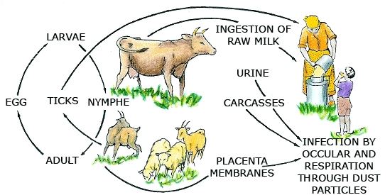
First described as transmitted by goat’s milk, is also a member of the swirl of the raving mad. Its first name comes from the Island of Malta , where it appeared early in the nineteenth century. It is a remarkable disease considering its geographical distribution. It is found in the Middle East, Africa and all over Europe. Its rapid evolution is characteristic. Starting with fever, any organ may become involved and lead to severe complications, just as in tuberculosis, which it seems to copy.

